Cryoablation, also called cryotherapy, is defined as therapeutic tissue destruction by freezing. It is a minimally invasive (percutaneous) thermal ablation technique that can treat kidney, bone, liver, lung, adrenal, and prostate lesions. It is particularly useful in treating tumors near critical structures. The visibility of the iceball intra-procedurally under MRI-guidance is a significant advantage of this technique.
Cryoablation in AMIGO is performed using an argon gas-based system (CryoHit, Galil Medical, Arden Hills, MN). Under MRI-guidance, 17-gauge cryoablation probes are placed by an attending interventional radiologist with the assistance of a clinical fellow-in-training. MRI scans are performed during each of four phases of the procedure: planning, targeting, monitoring, and survey.
MRI Guided Cryoablation Workflow in AMIGO
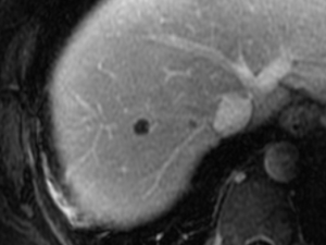
Planning
An initial planning MRI scan of the tumor is obtained. This scan allows the interventional radiologist to choose how many probes will be used and where to enter.
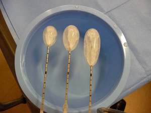
Planning
The number and positions of the probes are chosen based on the size of the tumor so that the resultant iceball will encompass the tumor plus a minimum 5-10 mm margin all around. Here, you can see different probes and their iceball sizes. No argon leaves the probe; it is confined in the probe and becomes extremely cold so as to generate the iceball.
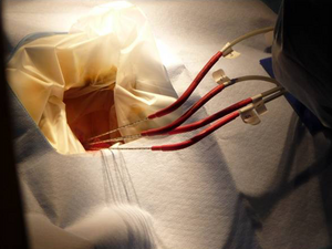
Targeting
Targeting phase of placing four probes. Freezing begins only after probes are placed in ideal locations using MR guidance.
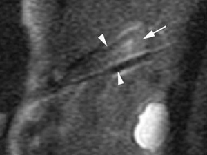
Targeting
Multiple MRI scans are taken during the targeting phase to assure proper location of the probes in the tumor and with the cryoprobes, ideally, approximately 1.5 cm apart.
Monitoring
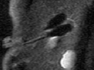
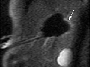
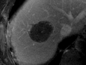
In the monitoring phase, cryoablation probes are activated simultaneously. The iceball generated during cryoablation will appear as a signal void (black in color) under MRI-guidance. Monitoring is used to confirm that the iceball has encompassed the tumor plus a margin. If necessary, a lower power can be used to prevent damage to adjacent critical structures.
Monitoring
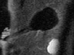
Iceball formation. The cryoablation protocol consists of a 15-minute freeze (argon gas), followed by a 10-minute thaw (helium gas), followed by a second 15-minute freeze.
Monitoring
1) Intracellular ice → Cell membrane rupture
2) Extracellular ice → Cellular dehydration (thaw)
3) Vascular stasis → Local tissue ischemia.
Formation
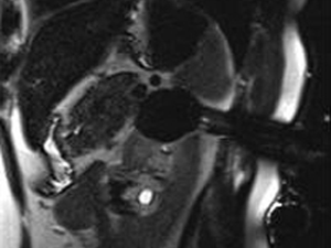
Iceball is monitored by MRI scans. Scans are made every 3 minutes during each freeze to watch the iceball grow and assess nearby critical structures.
Formation
Upon completion of the cryoablation protocol, the probes are removed and a final MRI scan acquired to survey the ablation site for possible complications.
Recent Publications
- Lee TC, Guenette JP, Moses ZB, Lee JW, Annino DJ, Chi JH. MRI and CT Guided Cryoablation for Intracranial Extension of Malignancies along the Trigeminal Nerve. J Neurol Surg B Skull Base. 2020;81 (5) :511-4. PMID: 33134018; PMCID: PMC7591355.
- Shono N, Ninni B, King F, Kato T, Tokuda J, Fujimoto T, Tuncali K, Hata N. Simulated Accuracy Assessment of Small Footprint Body-mounted Probe Alignment Device for MRI-guided Cryotherapy of Abdominal Lesions. Med Phys. 2020;47 (6) :2337-49. PMID: 32141080; PMCID: PMC7889307.
- Tokuda J, Wang Q, Tuncali K, Seethamraju RT, Tempany CM, Schmidt EJ. Temperature-Sensitive Frozen-Tissue Imaging for Cryoablation Monitoring Using STIR-UTE MRI. Invest Radiol. 2020;55 (5) :310-7. PMID: 31977600; PMCID: PMC7145748.
- Tokuda J, Chauvin L, Ninni B, Kato T, King F, Tuncali K, Hata N. Motion Compensation for MRI-compatible Patient-mounted Needle Guide Device: Estimation of Targeting Accuracy in MRI-guided Kidney Cryoablations. Phys Med Biol. 2018;63 (8) :085010. PMID: 29546845; PMCID: PMC5899055.
- Glazer DI, Tatli S, Shyn PB, Vangel MG, Tuncali K, Silverman SG. Percutaneous Image-Guided Cryoablation of Hepatic Tumors: Single-Center Experience with Intermediate to Long-Term Outcomes. AJR Am J Roentgenol. 2017;209 (6) :1381-9. PMID: 28952807; PMCID: PMC5698169.
- Liu X, Tuncali K, Wells III WM, Zientara GP. Automatic Iceball Segmentation with Adapted Shape Priors for MRI-guided Cryoablation. J Magn Reson Imaging. 2015;41 (2) :517-24. PMID: 24338961; PMCID: PMC4128910.
- Fairchild AH, Tatli S, Dunne RM, Shyn PB, Tuncali K, Silverman SG. Percutaneous Cryoablation of Hepatic Tumors Adjacent to the Gallbladder: Assessment of Safety and Effectiveness. J Vasc Interv Radiol. 2014;25 (9) :1449-55. PMID: 24906627.
- Dunne RM, Shyn PB, Sung JC, Tatli S, Morrison PR, Catalano PJ, Silverman SG. Percutaneous Treatment of Hepatocellular Carcinoma in Patients with Cirrhosis: A Comparison of the Safety of Cryoablation and Radiofrequency Ablation. Eur J Radiol. 2014;83 (4) :632-8. PMID: 24529593.
- Aghayev A, Tatli S. The use of Cryoablation in Treating Liver Tumors. Expert Rev Med Devices. 2014;11 (1) :41-52. PMID: 24308738.