Ciprian Catana, MD, PHD Director of Integrated PET/MR Imaging Athinoula…
Sila Kurugol, PhD: Advancements in Abdominal MRI: Enhancing Quantitative Imaging with Innovations in Diffusion-Weighted and Dynamic Contrast-Enhanced MRI

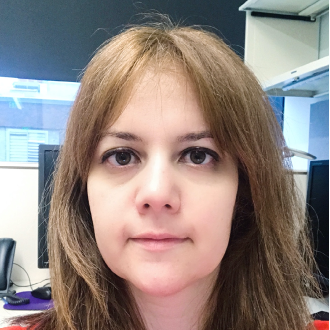
Sila Kurugol, PhD
Assistant Professor of Radiology
Director of Quantitative Intelligent Imaging Lab
Department of Radiology, Boston Children’s Hospital and Harvard Medical
Abstract
The presentation will highlight recent advancements in two key MRI sequences for abdominal imaging: Diffusion-weighted MRI (DW-MRI) and Dynamic Contrast-Enhanced MRI (DCE-MRI).
DW-MRI shows great promise in evaluating abdominal diseases, including cancer and noncancerous conditions in the liver, kidneys, and other organs. It is utilized for identifying tumors and abscesses, distinguishing between benign and malignant tumors, and pinpointing inflamed bowel segments in pediatric inflammatory bowel disease. However, abdominal diffusion MRI faces technical challenges, such as physiological motion (respiratory and peristaltic), image distortions, and a low signal-to-noise ratio. These challenges impact the reliability of quantitatively mapping diffusion parameters crucial for assessing tissue microstructure and clinical outcomes. To address these challenges, we’ve developed innovative techniques, including motion correction, self-supervised denoising, and distortion correction in the presence of motion. These advancements have the potential to improve the reliability of quantitative diffusion MRI in abdominal imaging.
Dynamic Contrast-Enhanced MRI (DCE-MRI) is essential in abdominal MRI, used for identifying and characterizing mass lesions and assessing renal function, specifically measuring glomerular filtration rate and differential renal function. However, 3D DCE-MRI encounters challenges, notably compromised spatiotemporal resolution and motion artifacts. The limitation in spatiotemporal resolution is due to rapid contrast dynamics lasting only a few minutes and relatively slow data acquisition for each temporal phase. This challenge is more pronounced in pediatric DCE-MRI due to smaller anatomical structures requiring higher spatial resolution and faster hemodynamics. We’ve developed reconstruction techniques based on compressed sensing and deep learning to enhance spatiotemporal resolution and facilitate accurate functional analysis. Additionally, we’ve introduced novel techniques for imaging infants and children, incorporating bulk motion detection and motion-compensated reconstruction. Moreover, we’ve implemented innovative approaches for respiratory motion correction, utilizing both prospective and retrospective motion techniques with various navigators such as pilot tone and FID navigators.
Short Bio
Dr. Kurugol serves as an Assistant Professor of Radiology at Harvard Medical School and leads the Quantitative Intelligent Imaging (QUIN) Research Group within the Computational Radiology Laboratory (CRL) at Boston Children’s Hospital. She holds degrees in Electrical and Computer Engineering—BS, MSc, and PhD—with her doctoral research focusing on pioneering machine learning techniques for medical image analysis. Following her PhD, Dr. Kurugol pursued postdoctoral research at Brigham and Women’s Hospital from 2011 to 2014, in Surgical Planning Lab (SPL). Subsequently, she joined the Computational Radiology Laboratory (CRL) at Boston Children’s Hospital, working under the guidance of Dr. Simon K. Warfield. During this time, she broadened her expertise in medical imaging, particularly MRI, to address practical clinical challenges. Establishing her own research group within the Radiology department at BCH, Dr. Kurugol’s recent work concentrates on advancing MR imaging techniques, developing motion correction methods, employing computational modeling strategies, and creating machine learning algorithms for image reconstruction, image enhancement and artifact reduction and image analysis. Her research aims to enhance the understanding and interpretation of medical images, focusing on deriving quantitative and functional biomarkers for the automatic assessment of various conditions., automated diagnosis of disease. Dr. Kurugol’s contributions have been recognized with several awards, including summa and magna cum laude awards from ISMRM and the Caffey Award from SPR, and best paper awards from IEEE conferences and workshops.